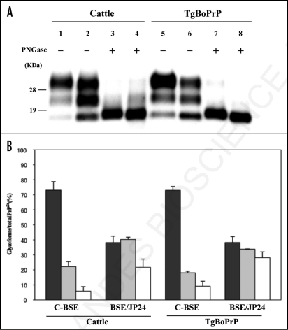
Figure 1.
Western blotting analysis of PrPSc from C-BSE and BSE/JP24. (A) The fragment of PrPSc in the cattle brain of C-BSE (lanes 1 and 3) and BSE/JP24 (lanes 2 and 4). PrPSc in the brain of TgBoPrP inoculated with C-BSE prion (lanes 5 and 7) and BSE/JP24 prion (lanes 6 and 8). All samples were digested with 50 µg/ml PK at 37°C for 1 h, and then samples in lanes 3, 4, 7 and 8 were treated with PNGaseF. PrPSc was detected by using mAb 6H4. Molecular markers are shown on the left (kDa). (B) The relative amount of the di-, mono- and non-glycosylated form of PrPSc in the C-BSE and BSE/JP24 prion affected individual. The results are mean ± standard deviation in five experiments. Bar diagram: diglycosylated form (black), monoglycosylated form (grey) and nonglycosylated form (white).

