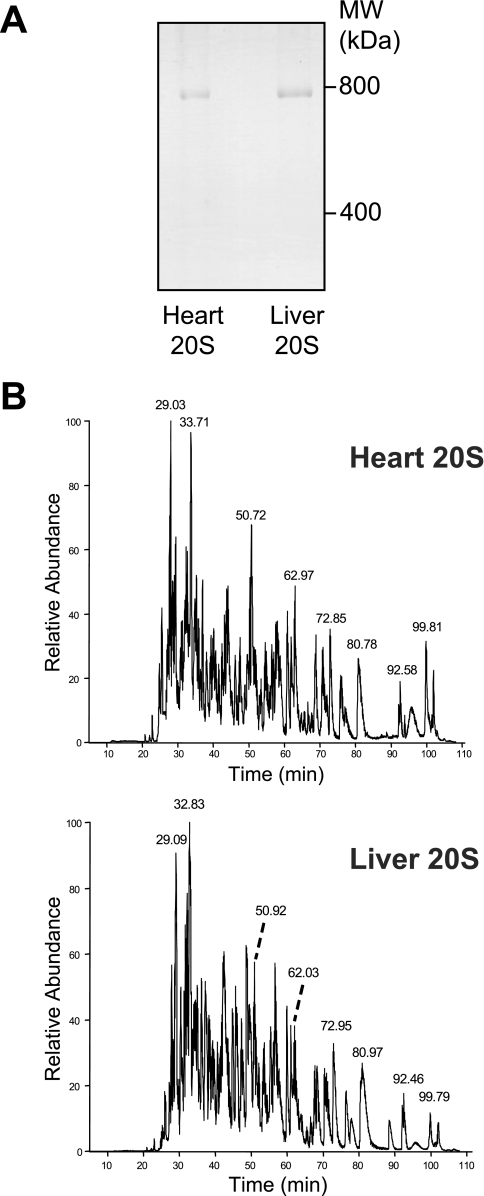
Fig. 1.
Analysis of purified murine cardiac and hepatic 20 S proteasomes. A, blue-native gel of purified murine cardiac and hepatic 20 S proteasomes. B, liquid chromatography of trypsin-digested heart and liver 20 S proteasome bands. The chromatograms for trypsin cleaved heart and liver 20 S proteasomes were similar, but distinct. Some of the major peaks in both samples are labeled.

