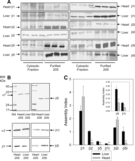
Fig. 3.
Comparison of free and assembled 20 S proteasome subunits in heart and liver by immunoblotting. A, comparison of constitutive and inducible proteasomes subunits in cytosolic fractions and purified 20 S from the heart and liver. Each lane contained 25 μg of cytosolic fraction or 1 μg of purified proteasome. B, comparison of proteasomes subunits in cytosolic fractions and purified 20 S from the heart and liver. Heart and liver 20 S proteasomes were run on SDS-PAGE, transferred to nitrocellulose, and probed with anti-proteasome antibodies. Each lane contained 1 μg of heart proteasome or 1 μg of liver proteasome. C, the assembly index for proteasome subunits in heart and liver cytosolic fractions. The assembly index is the ratio of the total amount of a proteasome subunit in the cytosolic fraction (free + partially assembled + assembled subunits) versus the amount of that proteasome subunit in the 20 S proteasome purified from the cytosolic fraction (only assembled subunits). The insert in Fig. 3C is an enlarged view of the assembly index for β5 and β1i.

