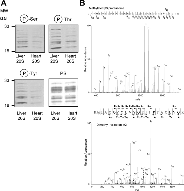
Fig. 4.
Comparison of post-translational modifications on purified 20 S proteasomes from heart and liver. A, comparison of the phosphoproteome of purified 20 S proteasomes from heart and liver. Each lane contained 2 μg of heart proteasome or 2 μg of liver proteasome. B, upper panel, mass spectra of a peptide from heart β6 proteasome subunit, which is mono-methylated on arginine; lower panel, a peptide from liver α2, which is dimethylated on lysine. # represents methylated arginine residue. The methylated peptide (β6) had an Xcorr of 4.114 and a precursor ion m/z of 1157.30 (3+ charge). @ represents dimethylated lysine residue. The dimethylated peptide (α2) had an Xcorr of 5.266 and a precursor ion m/z of 893.23 (3+ charge). Protein loading was controlled by Ponceau S stain (PS).

