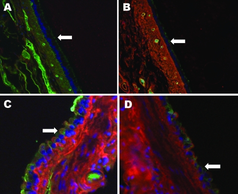
Figure.
Raccoon respiratory tissues stained with lectins specific for sialic acids (SAs) with α2,6- and α2,3-linkages. A) Upper trachea; B) lower trachea; C) bronchus; D) bronchiole. Arrows indicate endothelial lining of the tissues indicated. Green staining shows a reaction with fluorescein isothiocyanate–labeled Sambucus nigra lectin, which indicates SAs linked to galactose by an α2,6-linkage (SAα2,6Gal). Red staining shows a reaction with biotinylated Maackia amurensis lectin (detected with Alexa Fluor 594–conjugated streptavidin), which indicates an SAα2,3Gal linkage. Tissues were counterstained with 4,6,-diamidino-2-phenylindole dihydrochloride. Original magnification ×40 in panels A, B, and D and ×100 in panel C.

