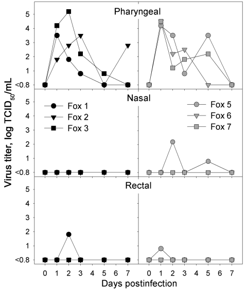
Figure 1.
Infectious virus titers obtained from pharyngeal, nasal, and rectal swabs of foxes infected intratracheally with highly pathogenic avian influenza (HPAI) virus (H5N1) (left, black symbols) or fed chicks infected with HPAI virus (H5N1) (right, gray symbols) at various time points after infection. No virus was isolated from any swabs of the negative-control foxes. TCID50, median tissue culture infectious dose.

