
Figure 4.
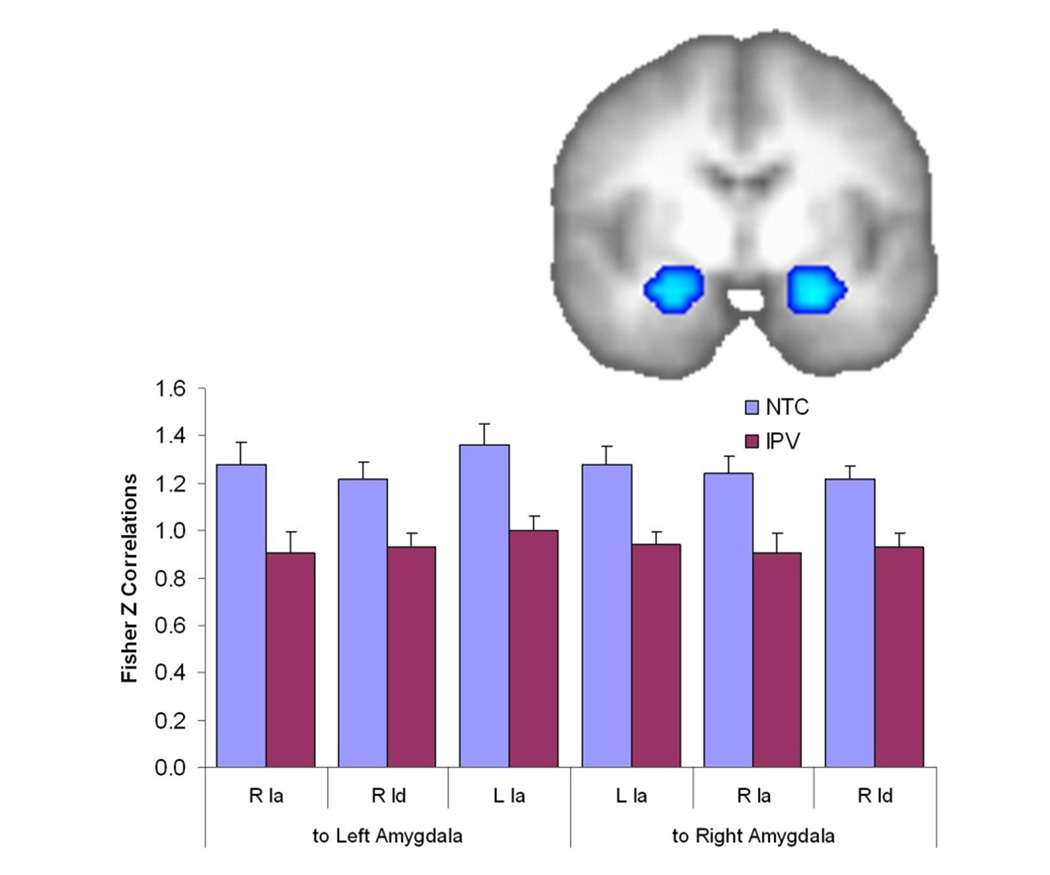
Reduced Functional Connectivity from the anterior insula (Ia), middle insula (Id), and posterior insula (Ig) to the Amygdala in the IPV group. ROI image shows the regions in the amygdala of significantly reduced functional connectivity from a left anterior insula seed; functional connectivity from the other insula regions is very similar to that displayed in the graph.