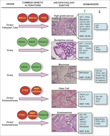
Figure 1. Histological and Molecular Heterogeneity in Epithelial Ovarian Cancers.
Common genetic alterations vary between different epithelial ovarian cancer sub-types. Highlighted in red are genes/pathways commonly inactivated in tumours; highlighted in green are genes commonly activated or amplified in epithelial ovarian cancer tumour specimens. Hematoxylin and eosin stained sections show typical histological and architectural appearance of the high-grade serous, borderline serous, mucinous, clear cell, and endometrioid sub-types. Biomarkers listed are those found in the study by Huntsman and colleagues to be highly expressed (i.e., samples positive in over 60% of tumours, green arrow), or lowly expressed (red arrow) in each histological sub-type. Median Ki67 labelling indices (a measure of the proportion of proliferating cells in a tumour sample) are given in bold type.

