
Figure 6. Spatial expression of bantam miRNA in developing eye disc and its modulation of rbf-induced apoptosis.
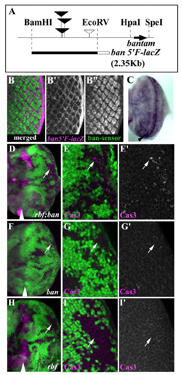
(A) Schematic representation of bantam flanking sequence. Bantam hairpin coding region is shown by a black arrow. Filled inverted triangles indicate P-element insertion sites that disrupted bantam function. An open inverted triangle indicates P-element insertion site associated non-bantam mutation. The 2.35Kb BamHI and EcoRV fragment was used to generate ban-5’F-lacZ shown at the bottom (filled black bar). The open box represents the LacZ reporter. β-gal expression from ban5’F-lacZ (magenta) and GFP expression from ban-sensor-GFP (green) are complementary (B-B”). High bantam expression is seen in interommatidial cells (β-gal, B’) while high bantam sensor is seen in photoreceptor cells (GFP, B”). Merged image is shown in (B). (C) In situ hybridization with DIG labeled lacZ RNA probe to ban5’F-lacZ eye disc. High level of expression in the MF and posterior interommatidial cells were seen. The MF groove indicates black arrowhead. (D-I’) bantam miRNA attenuates rbf induced cell death. Low (D) and high (E, E’) magnification images of rbf; ban double mutant cells showed increased Cas3 staining (magenta) in posterior mutant clones. Low (F) and high magnification (G, G’) images of bantam mutant clones showed background level of Cas3 staining. Low (H) and high magnification (I, I’) images of rbf mutant clones showed very low Cas3 labeling in the posterior, while clones spanning the MF showed extensive Cas3 labeling.