
Figure 1. Parietal cells are a key cellular component of the gastric epithelial progenitor niche.
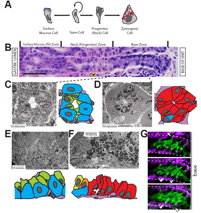
A, schematic of surface mucous and zymogenic lineage differentiation from a stem cell. The parietal and enteroendocrine lineages are not depicted. B, gastric unit stained with H&E. The unit opens out into the gastric lumen to the left; the lamina propria and muscle are to the right. Gastric unit zones are delineated above the image. The cells depicted in panel A are aligned with the zones in B in which they reside. The large eosinophilic (pink) cells are parietal cells (PCs, e.g., arrowhead). Scale bar = 50 μM. C-F, tEM images of gastric unit zones with accompanying cartoon traces of the EM image. PCs, blue; NCs, green; neck-to-zymogenic transitional (Ramsey et al., 2007), yellow/orange; ZCs, red; mesenchymal/endothelial cells, pink. C, cross section through the neck zone. Note how NCs (green) line the lumen apically but have scant basement membrane contact. D, cross section through the base zone. E-F, longitudinal sections through the progenitor (E) and base (F) zones. Note how the progression from NC to ZC involves not only a switch from electron lucent (mucous) to electron dense (zymogenic) granules but also substantial expansion of contact with the basement membrane. G, three serial slices from two-photon, 3-dimensional imaging of an entire gastric unit. The neck zone is shown, with NCs in green and PCs in purple. Note the thin NC projections (arrowheads) towards the basement membrane (dashed white line).