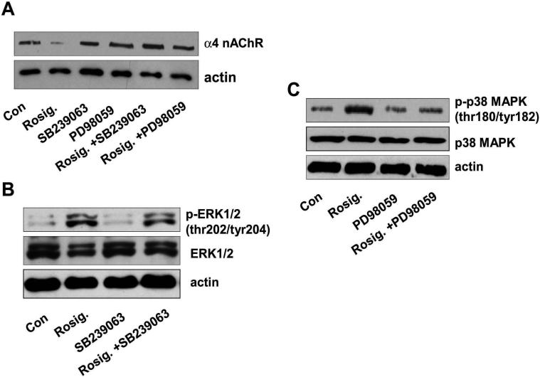
Figure 3. The inhibitors of p38 MAPK and ERK block the effect of rosiglitazone on expression of α4 nAChR.
A, Cellular protein (20 μg) was isolated from H1838 cells were treated with SB239023 (10 μM) or PD98059 (25 μM) for 2 h before exposure of the cells to rosiglitazone (Rosig.) for an additional 24 h. Afterwards, Western blot analysis was performed to detect the α4 nAChR protein. Actin was used for loading control for normalization purpose. Con, indicates untreated control cells. B, Cellular protein was isolated from H1838 cells treated with SB239023 (10 μM) for 1 h before exposure of the cells to rosiglitazone (Rosig.) for an additional 2 h. Afterwards, Western blot analysis was performed to detect the total ERK1/2 and phosphor-ERK1/2. Actin was used for loading control for normalization purpose. Con, indicates untreated control cells. C, Cellular protein was isolated from H1838 cells treated with PD98059 (25 μM) for 1 h before exposure of the cells to rosiglitazone (Rosig.) for an additional 2 h. Afterwards, Western blot analysis was performed to detect the total p38 MAPK and phosphor-p38 MAPK. Actin was used for loading control for normalization purpose. Con, indicates untreated control cells.

