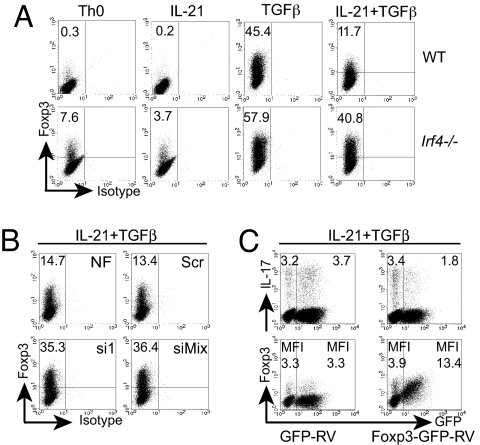
Fig. 5.
IRF4 is important for IL-21-mediated Foxp3 suppression. Flow cytometry of naïve Irf4−/− or WT CD4+ T cells differentiated as described in Fig. 1A and stained for intracellular Foxp3 (A) and of CD4+ WT cells nucleofected, cultured as described in Fig. 2B, and stained for Foxp3 (B). (A and B) Frequencies of Foxp3+ cells are indicated. (C) CD4+ T WT cells transduced with retroviruses expressing Foxp3-GFP or GFP, treated with IL-21 plus TGFβ, and stained for intracellular IL-17 or Foxp3. (Top) The percentages of GFP−IL-17+ (Left) or GFP+IL-17+ cells (Right) or the mean fluorescence intensity (MFI) of GFP− or GFP+ cells stained for Foxp3 are indicated. Representative data from three experiments are shown.

