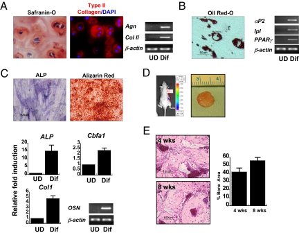
Fig. 2.
Developmental potential of hESCd-MSCs. (A) Chondrogenic differentiation in RGD-modified PEG hydrogels with TGF-β1 and ascorbic acid. Safranin-O staining (red) for negatively charged proteoglycans and type II collagen (type II collagen = red, nucleus = blue) in the pericellular signifies cartilaginous matrix production. RT-PCR confirmed chondrogenic differentiation of hESCd-MSCs with up-regulation of aggrecan and type II collagen. (B) The clusters of lipid-containing adipocytes were also detected by Oil red-O staining within 3 weeks of culture in adipogenic medium. Adipogenic differentiation was confirmed by the expression of αP2, lpl, and PPAR-γ by RT-PCR. (C) Bone-specific ALP was detected after 7 days, and mineralized deposits were evident with the alizarin red staining after 2 weeks of culture in osteo-inductive conditions. Osteogenic differentiation was confirmed by up-regulation of ALP, Cbfa1, and type I collagen by real-time PCR, and the expression of osteocalcin was confirmed by RT-PCR. (D) Gross image of in vivo-engineered bone tissues after 8 weeks of implantation. Microvascular perfusion and implant survival was assessed by luciferase detection of recombinant lentiviral vector-transduced hESCd-MSCs after 8 weeks of implantation. (E) H&E staining of the implanted scaffolds after 4 and 8 weeks. Histomophometric assessments showed increased bone area from 4 to 8 weeks (n = 4). UD, undifferentiated hESCd-MSCs; Dif, differentiated hESCd-MSCs.

