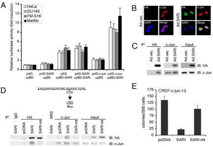
Fig. 5.
SARI directly interacts with c-JUN. (A) Mammalian two-hybrid assay was performed using pAD-c-jun and pBD-SARI in HeLa, DU145, FM516-SV, and MeWo cells as described in Materials and Methods. Luciferase activity was normalized by β-galactosidase activity. The data represent mean ± SD of three independent experiments. (B) HeLa cells were infected with Ad.vec or Ad.SARI at 50 pfu/cell. The cells were fixed 24 h later and subjected to immunofluorescence analysis using anti-HA and anti-c-JUN primary antibodies and FITC- and rhodamine-conjugated secondary antibodies, respectively. Mounting medium containing DAPI was used for staining the nucleus. Confocal laser scanning microscopy was used to analyze the images. (C) HeLa cells were infected with Ad.vec or Ad.SARI at 50 pfu/cell. Total cell lysates were used for co-immunoprecipitation analysis using anti-HA antibody for immunoprecipitation and anti-c-JUN antibody for immunoblotting and vice versa. (D) Expression plasmid in which Leucine59 of SARI was mutated to proline was generated (SARI-mt). HeLa cells were transfected with empty vector (pcDNA) or expression plasmids expressing WT SARI or SARI-mt. The cell lysates were subjected to co-immunoprecipitation analysis as described in C. (E) CREF-c-jun-13 cells were transfected with empty vector (pcDNA) or expression plasmids expressing WT SARI or SARI-mt and subjected to clonogenic assay for 2 weeks upon selection with hygromycin. Data represent mean ± SD of three independent experiments.

