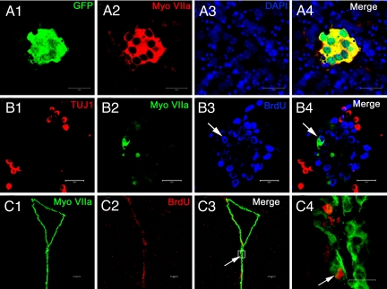
Fig. 1.
In vivo and in vitro proliferation of ependymal cells. (A1–4) Adult NSCs were isolated from the lateral wall of the LV (obtained from Myosin VIIA-GFP mice). Dissociated cells proliferated into neurospheres. Within the neurosphere a small cell colony was colabeled with GFP (A1) and myosin VIIA (A2), indicating the possible proliferation of myosin VIIA positive cells. Nuclei were labeled with DAPI (A3 and 4). (B1–4) After the neurospheres attached to the coverslips, some of the progenies expressed the early neuronal maker β tubulin III (TUJ1) (B1) and myosin VIIA (B2), respectively. Newly generated cells were labeled with BrdU. Arrows indicate an in vitro proliferated cell, which was simultaneously labeled with myosin VIIA and BrdU (B3 and 4). (C1–4) Cryosection of an adult brain taken from a BrdU treated mouse. The ependymal layer of the LV was clearly and specifically labeled with myosin VIIA (C1). Nuclei of proliferated cells were labeled with BrdU (C2). Arrows indicate an ependymal cell that was colabeled with myosin VIIA and BrdU. Higher magnification of the colabeled ependymal cell within the box area can be found in panel (C4), which indicates the possible in vivo proliferation. (Scale bars: A1–B4 and C1–3, 100 μm.)

