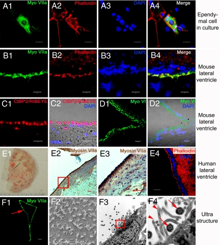
Fig. 2.
In vitro and in vivo structural profile of adult ependymal cells. (A1–4) Cultured adult ependymal cells remain myosin VIIA-positive (A1). Phalloidin-labeled actin-rich sterocilia-like appendages were found on the apical surface of the cells (A2). (B1–D2) Cryosection of the lateral wall of the LV of adult mice. Ependymal cells were clearly and specifically labeled with myosin VIIA (B1) and phalloidin (B2–4). Hair cell synaptic protein CtBP2/RIBEYE was observed in ependymal cells of the LV (C1 and 2). The ependymal cell layer of the LV also expressed myosin VI, an early HC marker (D1 and 2). Myosin VI-positive cells are shown in green (D1), whereas the nuclei-stain is in blue. The light microscope and merged image are represented in (D2). (E1–4) Myosin VIIA was expressed in adult human ependymal cells. Panel (E1) is a photomicrograph of a normal adult human brain sliced and frozen within 18 h of death. Postmortem, a wedge around the LV region was taken from the brain then fixed, sectioned and stained to provide panels (E2–4). The red dashed lines indicate the LV region (E1). Adult human ependymal cells also expressed myosin VIIA (E2). The boxed area of panel (E2) is enlarged in panel (E3) to demonstrate myosin VIIA expression. Phalloidin-labeled actin stereocilia-like appendages were found on the apical aspects of human ependymal cells (E4). Scanning (F2) and transmission electron microscopy (F3) of the lateral ventricle region (red arrow in F1) demonstrated that ependymal cells are also equipped with structural profiles of stereocilia and kinocilia, similar to HCs. The boxed area of panel (F3) was enlarged in panel (F4), showing stereociliary appendages (arrow heads) and the characteristic 9 + 2 microtuble structure of kinocilia (arrows). The nuclei were labeled with DAPI. (A3, A4, B3, B4, C2, D2, and E4). (Scale bars: A1–D2, E4, and F2, 20 μm; F1 and E2 and 3, 100 μm.)

