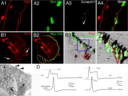
Fig. 5.
NSC-derived neurons demonstrated defining characteristics of SGNs. (A1–4) Adult NSCs were cocultured with HCs. Nerve endings of a NSC-derived neuron in contact with a cocultured HC. The accumulation of synapsin 1 at the nerve ending suggests that the contact between NSC-derived neurons and HCs may develop into a real synapse. (B1–4) NSC-derived neurons also established synapse-like contacts with HCs at the organ level. The organ of Corti was collected from P3 mice and SGNs were removed. To eliminate the residual SGNs, the dissected organ of Corti was treated with β-bungarotoxin (0.5 μM) for 48 h then cocultured with NSCs. Neuronal cells were labeled with TUJ1 and HCs were labeled with myosin VIIA. As panel (B1) demonstrates, residual SGNs were selectively removed from the organ of Corti after pretreatment with β-bungarotoxin. Nerve fibers of NSC-derived neurons (arrows) penetrated the organ of Corti and established contacts with HCs (B2). The dashed line marks region in panel (B2) that was enlarged and reconstructed into a 3-D image in panel (B3); a cluster of NSC-derived neurons projected fibers into the organ of Corti and integrated with HCs. The accumulation of synapsin 1 indicates that the contacts between HCs and NSC-derived neurons may develop into synapses. The boxed area in panel (B3) was further enlarged in panel (B4) to show the synapse-like contacts. (C) Ultra structure of the nerve endings of a NSC derived-neuron that contacted cocultured HCs (arrowheads indicate the stereocilia). Synaptic vesicles were found within the nerve ending (arrow). (D, left panel) Simultaneous current-clamp recordings from a HC and a NSC-derived neuron in close contact. The HC was injected with 0.7-nA positive current. The NSC-derived neuron was injected with a sustained negative current to establish a membrane potential of −83 mV. Under these conditions, sufficient depolarization of the HC elicited action potentials in the NSC-derived neuron. (D, right panel) Similar results could be seen by a HC depolarization of 0.8 nA, leading to a corresponding action potential from a SGN. For SGNs in culture, action potentials could be generated at −50 mV resting potential. (Scale bars: A1–4, B3, C1–4, 20 μm; B1 and 2, 100 μm.)

