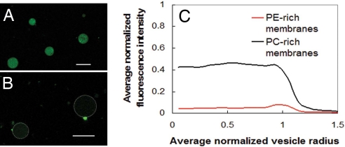
Fig. 1.
The limitation of dye leakage experiments. (A) Alexa480 fluorescence intensity from tagged dextran encapsulated in antimicrobial-treated DOPG/DOPC = 20/80 GUVs. (B) Alexa480 fluorescence intensity from tagged dextran encapsulated in antimicrobial-treated DOPG/DOPE = 20/80 GUVs is extremely weak because of leakage. Dotted white lines indicate the outline of vesicle membranes. (Scale bars, 10 μm, in A and B.) These dye leakage results and others suggest that a trend of increasing leakage with increasing PE lipid content in membranes after treated with the phenylene ethynylene antimicrobials. (C) Confocal microscopy results show circularly integrated intensities of fluorescently tagged dextran inside antimicrobial treated GUVs for PE-rich membranes composed of DOPG/DOPE = 20/80 and for PC-rich membranes composed of DOPG/DOPC = 20/80. Data from 10 GUVs are averaged into the PE-rich trace and 17 into the PC-rich trace. Fluorescence intensity is normalized so that untreated vesicles have fluorescence intensity of 1.0, so clearly there is significant nonspecific leakage, which complicates interpretation.

