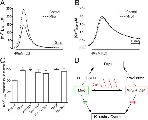
Fig. 6.
Effect of Miro on [Ca2+]c and [Ca2+]m signaling in neurons. (A and B) Miro1 over-expression increases 40 mM KCl-induced [Ca2+]m uptake. Cortical cultures were cotransfected with aequorin targeted to mitochondria (A) or cytosol (B) and Miro1WT and Ca2+ uptake was measured luminometrically as described in Materials and Methods. The [Ca2+]c peak was 1.97 ± 0.16 μM in control versus 1.87 ± 0.08 μM in Miro1-overexpressing neurons (n = 8, P = 0.579). (C) Changes in mitochondrial Ca2+ uptake induced by overexpression of Miro1 and Miro2 mutants. Data presented as % of control, from 3–6 independent experiment (n ≥ 15), **, P < 0.001 vs. control group. (D) Scheme illustrating the bidirectional Ca2+-dependent control of mitochondrial motility and fusion-fission dynamics by Miro proteins.

