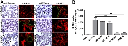
Fig. 5.
Immunization with BCG-M2 and BCG-N reduces virus presence in lung tissues after RSV infection. (A) Four days after infection, lungs were removed, fixed with paraformaldehyde, and stained with an HRP-labeled anti-RSV antibody [first and third columns (arrowheads show positive staining)] or with a biotin-labeled anti-F antibody followed by streptavidin-FITC (second and fourth columns), as described in SI Methods. Fluorescence counterstaining derives from a Cy3-conjugated anti-von Willebrand factor antibody. Positive staining is observed in lungs of unimmunized, UV-RSV, and WT-BCG-immunized mice. Data shown are representative of 3 to 6 independent experiments. (B) Total RNA from lungs of control and infected animals were obtained and reverse transcribed to quantify the number of N-RSV copies by real-time PCR. Data are expressed as the number of N-RSV gene copies per 5,000 copies of β-actin gene (**, P < 0.01, 1-way ANOVA).

