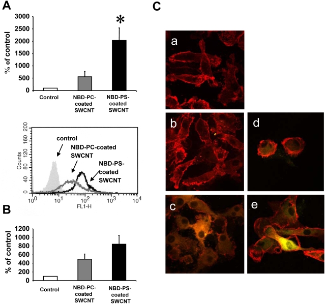
Figure 6. Primary human monocyte-derived macrophages and dendritic cells recognized SWCNT functionalized with PS but not with PC.
A. M-CSF-activated HMDM (0.5×10⁶ cells/ml) were incubated with NBD-PC- or NBD-PS-coated SWCNT (100 µg/ml) for 2 h and then subjected to flow cytometric evaluation of uptake of nanotubes. Macrophages incubated without SWCNT were included as an autofluorescence background control, and values are reported as percentage of background control. Representative histograms are shown below the bar graph. PS-coated SWCNT were observed to be taken up by HMDM to a higher degree than PC-coated SWCNT (p<0.002) (n = 4). B. MDDC (0.5×10⁶ cells/ml) exposed to NBD-PC- or NBD-PS-coated SWCNT (100 µg/ml) for 24 h at 37°C were assessed by flow cytometry. Data are reported as above. A tendency toward higher degree of uptake of PS-coated SWCNT was observed compared to PC-coated SWCNT in these cells (p<0.06) (n = 3), was seen for these cells. Similar results were obtained when uptake was monitored at 2 h (data not shown). C. Confocal microscopic imaging of MDDC incubated in the absence of SWCNT (a), or in the presence of NBD-PC-coated SWCNT for 2 h (b) or 24 h (d), or NBD-PS-coated SWCNT for 2 h (c) or 24 h (e), respectively. Counterstaining with antibodies to HLA-DR (red) was performed to visualize the plasma membrane of dendritic cells and the yellowish green color represents NBD-PS-coated SWCNT inside the cells. Original magnification - 63×2. Data are mean±s.d.

