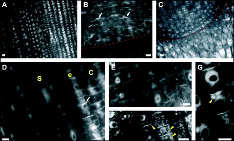
Figure 2.
IAA labelings in maize root apices: subcellular and cellular distributions. (A, B) In the untreated root, IAA enriched cross-walls (end-poles) are prominent in stele cells of the transition zone (A) and the whole quiescent centre (QC) (B). (C, F and G) In BFA-treated root tips (2 h), IAA labeling of end-poles vanishes in the stele while the nuclear labelling gets more prominent. BFA treatment shifts IAA signal into BFA-induced compartments. (D) In cells of the cortex, intensity of the synapse labelling gets weaker while labelling of nuclei increases. White arrowheads point on auxin-enriched BFA-induced compartments. Red Line in (B) and (C) marks the border between meristem and root cap. (S, stele; e, endodermis; C, cortex) Bars: 10 µM.

