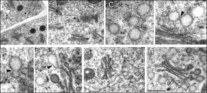
Figure 1.
Control cells of maize root epidermis. (A,B) Numerous Golgi stacks with loosely associated vesicles of trans/post-Golgi network (TGN/PGN) (boxed areas in B) and multivesicular bodies (MVBs, indicated by arrow in A). (C,D) Higher magnification views of TGN/PGN vesicles from boxed areas in part B reveal stalk-like connections (empty arrowheads) and bridge-like partial fusions (filled arrowheads). (E,F) Pear-shaped vesicles resulting from the advanced fusion between TGN/PGN vesicles (filled arrowheads). (G,H) Abundant TGN/PGN compartments near or at some distance from Golgi stacks (see also B and C). Arrowheads indicate bridge-like connections. Bar = 1.2 µm for A; 1 µm for B; 0.25 µm for C and F; 0.3 µm for D; 0.2 µm for E; 0.5 µm for G and 0.35 µm for H.

