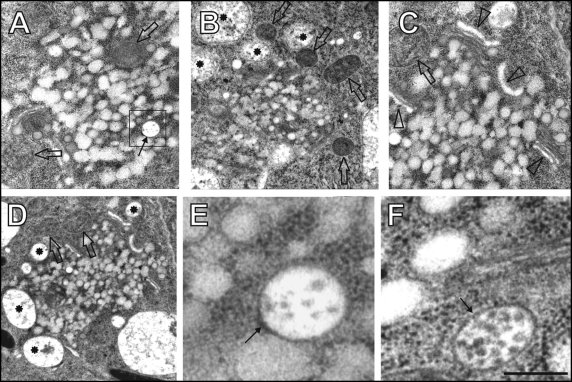
Figure 3.
60 minutes of brefeldin A (BFA) treatment. (A–D) Large BFA-induced compartments containing clusters of fused TGN/PGN vesicles and MVBs (arrow in A) as well as peripherally associated mitochondria (empty arrows), small vacuoles (stars), and Golgi stacks having inflated cis-cisternae (arrowheads),. Boxed area in A is presented as E. (E,F) Detail views on MVBs (arrows). Bar = 0.83 µm for A and C; 1.35 µm for B; 1.48 µm for D; 0.17 µm for E; 0.2 µm for F.

