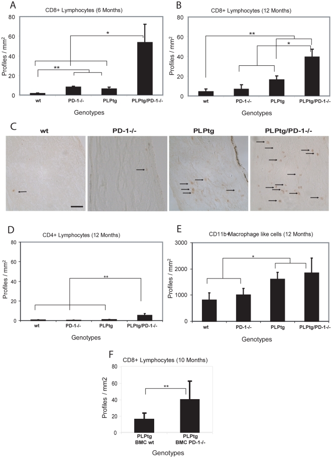
Figure 1. Quantification and immunohistochemical detection of CD8+ and CD4+ lymphocytes and CD11b+ macrophage-like cells in longitudinal sections of optic nerves of wt, PD-1-/-, PLPtg and PLPtg/PD-1-/- mice and of CD8+ cells in bone marrow chimeric mice.
- A, B. Quantification of CD8+ lymphocytes in 6 (A: n = 3) and 12 (B: n = 3–7) months old mice of different genotypes. C. Immunohistochemical detection of CD8+ lymphocytes in the optic nerve of 12 months old mice of different myelin and immunological genotypes. Arrows indicate positively labelled CD8+ lymphocytes. D. Quantification of CD4+ lymphocytes in 12 months old mice (n = 3–7). Note that in the myelin mutants, both CD8+ and the generally scarce CD4+ lymphocytes are substantially increased in the absence of PD-1. E. Quantification of CD11b+ macrophage-like cells in 12 months old mice (n = 3–6). Note that the number CD11b+ cells does not differ significantly in PLPtg/PD-1+/+ and PLPtg/PD-1-/- double mutants. Similar results are obtained in irradiated and non-irradiated (RAG-1-/-) PLPtg bone-marrow recipients. The rather small increase of CD11b+ cells is not surprising, since the molecule in focus (PD-1) is a component of predominantly T-lymphocytes rather than of macrophages/microglial cells. F. Quantification of CD8+ cells in the CNS of 10 months old PLPtg bone marrow chimeras (BMCs) which were transplanted with either wt or PD-1-/- bone marrow (n = 8–9). Note that also in bone marrow chimeras, CD8+ T-lymphocytes are substantially elevated in the CNS of PLPtg mutants in the absence of PD-1. Error bars represent standard deviations. * p- value<0.05, ** p - value≤0.01. Scale Bar: 50 µm.

