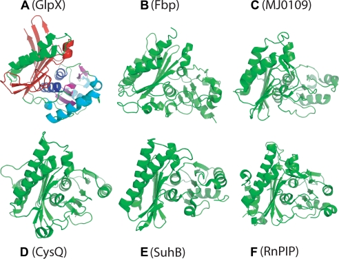
FIGURE 3.
Monomer structures of GlpX and other FBPases and Li+-sensitive phosphatases. A, E. coli GlpX (FBPase II; PDB code 3bih); B, E. coli Fbp (FBPase I; PDB code 2q8m); C, MJ0109 (FBpase IV; PDB code 1dk4); D, E. coli SuhB (IMPase; PDB code 2qfl); E, CysQ from B. thetaiotaomicron (PAPase; PDB code 3b8b); and F, RnPIP (PIPase; PDB code 1jp4). All of the polypeptides are shown in equivalent orientations revealing the presence of a five-layered α-β-α-β-α structure. The α/β layers of GlpX are shown in different colors: α1, green; β1, red; α2, blue; β2, magenta; α3, cyan.

