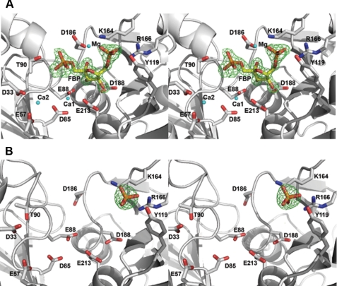
FIGURE 5.
Wide-eye stereo view of the GlpX active site. A, FBP bound in the GlpX-FBP complex (PDB code 3d1r); B, phosphate bound in the GlpX-phosphate complex (PDB code 3big). The ligand, metal ions (Mg, Ca1, and Ca2) and selected residues of GlpX (D61A) in contact with the ligand and metal ions (cyan spheres) are shown as a stick diagram along with a GlpX ribbon (gray). Difference electron density was calculated by removing the fructose 1,6-bisphosphate from the final model, followed by several rounds of maximum likelihood refinement using Refmac5. The resulting Fo - Fc map was contoured at 4.2σ.

