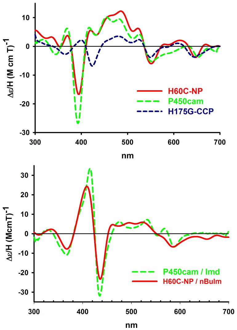
Figure 2.
MCD spectra of ligand free H60C_NP1 compared with the 5-coordinate thiolate complex of camphor bound P450cam, and bis-aquo coordinated H175G_CCP (upper panel) suggests that H60C_NP1 is coordinated by Cys-60. In the presence of nBuIm (one equivalent), the MCD of H60_NP1 is converted to a low spin complex (lower panel) that is similar to the mixed imidazole/thiolate complex of P450cam in the presence of imidazole. Samples were prepared in phosphate buffer, pH = 7.4 and 1 mM camphor and MCD spectra run at 4°C.

