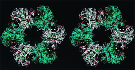
Figure 4.

Location of oxidized and intact methionine residues in glutamine synthetase. This stereo figure was created by the program rasmol (19) using the coordinates determined by Almassy et al. (18), deposited in the Brookhaven Data Base (reference 2GLS). For clarity, only one of the two hexamers is shown, and subunits are alternately colored blue and white. The sulfur groups of methionine residues are shown as balls, with intact residues in green and the oxidized residues in red. The active sites are formed by two adjacent subunits, and the Mn2+ in the core of the active sites is shown in yellow. The oxidizable methionine residues appear to form an array about the entrance to the active site bay.
