Fig. 1.
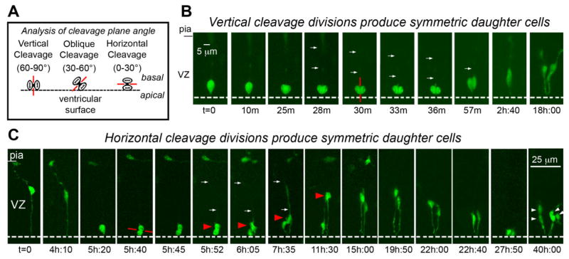
Radial glial (RG) cells undergo symmetric self-renewing divisions during early stages of cortical development. A: Classification of cleavage plane analysis. B: Time-lapse imaging of a single RG cell in an organotypic slice culture prepared from an embryonic day (E)13 rat. The RG cell is shown entering metaphase at the surface of the lateral ventricle at t = 0. The dotted white line indicates the surface of the lateral ventricle. A vertically oriented cleavage furrow (red line) begins developing t = 30 m, and can be clearly visualized at t = 33 m and 36 m. Both daughter cells acquired the morphology of RG cells and remained in the ventricular zone (VZ). C: Horizontal cleavage plane divisions at the ventricular surface also produce daughter cells with symmetric morphologies and behaviors. A single bipolar RG cell at E13 is shown in G1-phase at t = 0. The RG cell body rose slightly in the VZ before descending to the ventricular surface (t = 5h:20m). The pial fiber became very thin and faint during mitosis but was detected post hoc (see Suppl. Movie 2). The RG cell divided with a horizontal cleavage plane (red line) at t = 5h:40m. After division the soma of the basal daughter cell (red arrowhead) moved away from the ventricle at a faster rate than its apical sibling. This behavior might be interpreted to signify an asymmetric fate for the daughter cells. Nonetheless, extended time-lapse imaging demonstrated that both daughter cells retained contact with the ventricle, resumed IKM, and divided at the ventricular surface (t = 40 h). We therefore classified this RG division as symmetric self-renewing. An unrelated cell with the morphology of a tangentially migrating cell was present in the marginal zone of the viewing field from 5–11 hours. Time elapsed is shown in either minutes (m), or hours and minutes (hh:mm) as indicated below each sequence. The entire time-lapse sequences can be viewed in Supplemental Movies 1, 2. A magenta-green version of this figure can be viewed online as Supplementary Figure 1.

