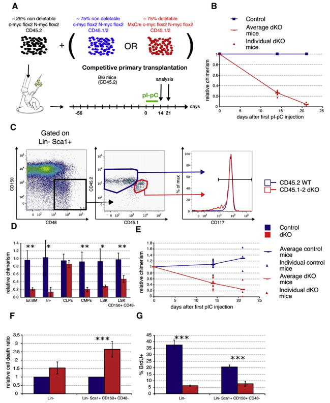
Figure 4. Bone Marrow Failure in dKO Mice Is Hematopoietic Autonomous, and dKO HSCs Show Decreased Proliferation and Survival.
(A) Competitive bone marrow chimeras. Mice were reconstituted with equal amounts of c-mycflox/flox;N-mycflox/flox cells and undeleted MxCre;c-mycflox/flox; N-mycflox/flox BM cells. After stable engraftment (8 weeks), pIpC was administered to the mice to induce deletion. The chimeras were analyzed 14 or 21 days after the first pIpC injection.
(B) Kinetics of loss of donor dKO cells in mixed chimeras. Results are mean ± SD. D14, n = 8; D21, n = 3.
(C) dKO HSCs present in mixed chimeras do not downregulate cKit (CD117) cell-surface expression. BM from competitive chimeras 14 days after pIpC treatment was analyzed and gated on Lin−Sca1+CD150+CD48−. The expression levels of cKit in the CD45.2+ (WT) rescue and in the CD45.1+/2+ (dKO) population were compared. Representative FACS plots from a total of six mice are shown.
(D) Degree of relative chimerism on progenitor and stem cell populations. CLPs (Lin−cKitintSca1int CD127+), CMPs (Lin−cKit+Sca1−); n > 3.
(E) Degree of relative chimerism of SLAM-HSCs. Triangles, individual mice; horizontal bar, the mean value for each genotype. D14, n = 8; D21, n = 3.
(F) Chimeras 14 days after the first pIpC treatment: BM was stained with stem cell markers, and apoptosis was assessed by TUNEL using FACS. Mean ± SD is shown; n = 3.
(G) Chimeras 14 days after the first pIpC treatment were administered BrdU for 15 hr before percent BrdU incorporation was assessed. Mean ± SD is shown; n = 3.

