
Figure 2.
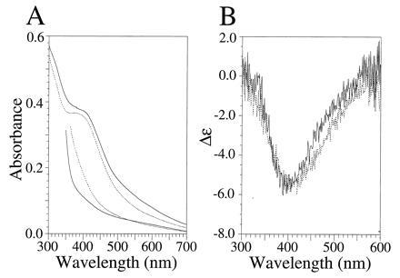
UV-vis and visible CD characterization of the [4Fe–4S] ferredoxin maquettes FdM and HLH–FdM. (A) UV-vis spectra of oxidized (upper traces) and dithionite-reduced (lower traces) [4Fe–4S] ferredoxin maquette (broken line) and HLH [4Fe–4S] ferredoxin maquette (solid line). Oxidized and reduced spectra were obtained from samples with equivalent peptide concentrations. The rising absorbance below 400 nm is due to dithionite. (B) Visible CD spectrum of the oxidized FdM (broken line) and HLH–FdM (solid line). All spectra contained 33 μM peptide in an anaerobic cuvette of 1.0-cm path length (10 mM KPi and 100 mM KCl, pH 8.0).