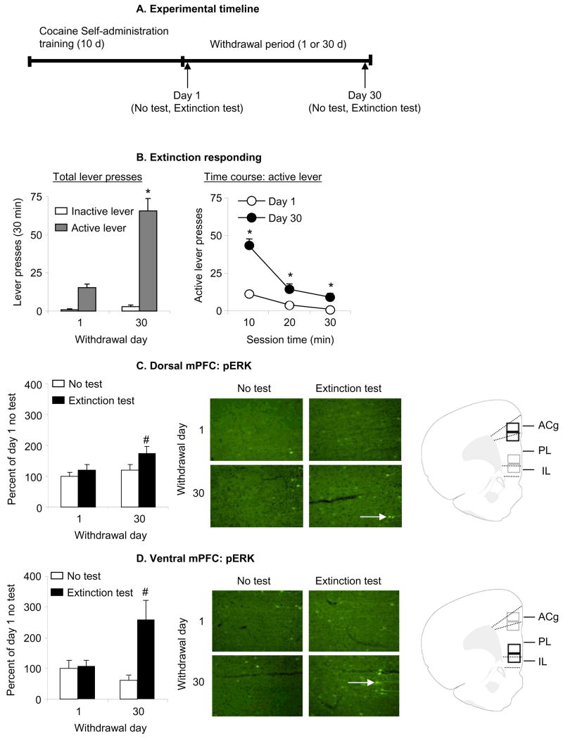
Figure 1. Effect of cue-induced cocaine seeking in extinction tests on p-ERK levels.
(A) Outline of experimental timeline. Four groups of rats self-administered cocaine for 6 h/d for 10 d. On withdrawal days 1 or 30, the rats were either tested or not tested for cocaine seeking in extinction tests, and their brains were removed for p-ERK analysis. (B) Test for cue-induced cocaine seeking: data are mean±sem number of lever-presses on the previously active lever and on the inactive lever during the tests for cocaine seeking performed under extinction conditions after 1 day or 30 days of withdrawal. During the test sessions, cocaine was not available and lever presses resulted in the delivery of the tone-light cue previously paired with cocaine. All rats were chronically housed in the self-administration chambers during training; the rats in the 30 days withdrawal groups were housed in their home cage in the animal facility after the self-administration training. Non-reinforced lever responding after 30 withdrawal days was higher than after 1 withdrawal day. * Different from Day 1, p<0.01, n = 9–11 per group). (C-D) p-ERK levels in dorsal or ventral mPFC on day 1 or 30 of withdrawal in the test or no test conditions; data are depicted as percent of mean Day 1 No Test condition. p-ERK positive cells are indicated with white arrows. Rats in the Extinction Test condition were trained to self-administer cocaine and were exposed to the cocaine cues in a 30 min extinction test after 1 or 30 withdrawal days. Rats in the No Test condition were trained to self-administer cocaine and were not exposed to the cocaine cues after 1 or 30 withdrawal days. Exposure to cocaine cues in the extinction tests increased the number of p-ERK positive cells (white arrows) in both dorsal and ventral mPFC after 30 days but not 1 day of withdrawal; this effect was more pronounced in ventral mPFC than in dorsal mPFC. Abbreviations: ACg, Anterior cingulate cortex; PL, Prelimbic cortex; IL, infralimbic cortex. # Different from the other 3 groups, p<0.05.

