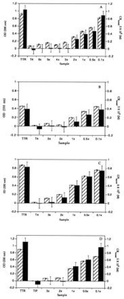
Figure 3.

Inhibition of TTR amyloid fibril formation by T4 and TIP. The extent of fibril formation in the presence and absence of inhibitor at pH 4.4 was probed at 72 h by an optical density measurement at 330 nm (hatched bars) and by a quantitative Congo red binding assay (solid bars). (A) Wild-type fibril formation. From left to right, fibril formation in the absence of T4 and in the presence of T4 alone (no TTR) and TTR fibril formation in the presence of the indicated equivalents of T4 (3 eq of T4 = 10.8 μM) are shown. Inhibition of (B) V-30-M and (C) L-55-P TTR by T4 at pH 5.0. (D) Inhibition of wild-type TTR by TIP. The labeling of the axes and the organization of the data in B–D is the same as that in A.
