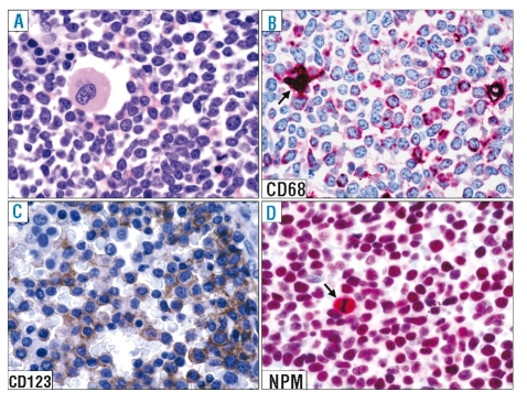
Figure 2.
Expression pattern of nucleophosmin (NPM) in BPDC neoplasm (bone marrow involvement). (A) Diffuse marrow infiltration by blast cells. A residual megakaryocyte is present (bone marrow biopsy; hematoxylineosin; ×800). (B) Leukemic cells express the macrophage-restricted form of the CD68 antigen with a dot-like pattern; the arrow indicates a histiocyte. (C) Most leukemic cells are strongly positive for the CD123 molecule. (D) Leukemic cells show a nucleus-restricted positivity for nucleophosmin which is indicative of wild-type NPM1 gene. A mitotic figure (arrow) shows the expected nucleophosmin cytoplasmic positivity. (B–D) Paraffin sections from bone marrow trephines immunostained with monoclonal antibodies against CD68 (PG-M1), CD123, and nucleophosmin (clone 376). (B) and (D), immunophosphatase alkaline (APAAP) technique; hematoxylin counterstain; ×800; (C): immunoperoxidase procedure hematoxylin counterstain; ×800.

