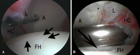
Fig. 1A–B.
Arthroscopic photographs of two different cases are shown. (A) In this arthroscopic photograph of a left hip, the hip capsule (HC) is to the left. The acetabulum (A) and labrum (L) are at the top of the photograph. Cartilage lesions (black arrows) are observed on the superior femoral head (FH) produced at the moment of the insertion of the spinal needle from the anterolateral portal. A probe is being introduced from the anterior portal on to the anterior acetabulum. (B) In an arthroscopy photograph of a left hip, the acetabulum (A) and labrum (L) are at the top of the photograph, and cartilage lesions caused by a cam impingement deformity are at the anterior rim (*). The hip capsule (HC) is to the right. A slotted cannula is being introduced through the anterior hip capsule. The black arrow points to a deep scuff on the femoral head (FH).

