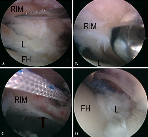
Fig. 4A–D.
A series of arthroscopic photographs taken from a right hip is shown. (A) A capsulotomy-capsulectomy of the anterior hip capsule has been performed. The labrum (L) has been detached from the acetabular rim (RIM) using an arthroscopic knife. The femoral head (FH) is at the bottom. (B) The acetabular rim (RIM) is being reshaped using an arthroscopic burr. The labrum (L) is at the bottom of the photograph. (C) A suture anchor has been introduced in the acetabular rim (RIM). The black arrow points to the articular on the anterior acetabular wall. The labrum is at the bottom of the photograph. (D) In this photograph, two anchors have been implanted and the labrum (L) reattached. Traction has been released. Recreation of the labral seal around the femoral head (FH) is demonstrated.

