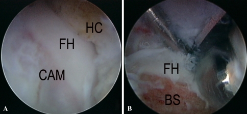
Fig. 6A–B.
Arthroscopic photographs of a right hip with a cam impingement deformity are shown. (A) The cam (CAM) deformity is observed at the femoral head (FH) and neck junction. The deformity has been exposed through a capsulectomy. The medial margin of the hip capsule (HC) is to the right. (B) The cam deformity has been reshaped using a burr. The burred surface (BS) is at the bottom. Articular cartilage from the femoral head (FH) is observed medial to the burred surface (BS). A probe is used to retract the hip capsule.

