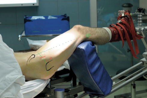
Fig. 1.
The patient is positioned supine with the traction device. The traction is only applied on the operated lower limb during the acetabular time. The approach is made with slight flexion. No fluoroscopy is needed. The incision is anterolateral and approximately 2 to 4 cm. The skin incision is parallel to the classic incision described by Hueter but moved downward approximately 1.5 cm to prevent injury of the lateral nerve of the thigh. The incision is centered on the summit of the great trochanter. Another more lateral and lower portal is used for the scope.

