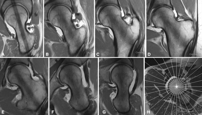
Fig. 6A–H.
(A) Example of a MRA radial slice at 3 o’clock position (posterior) at the femoral neck axis. (B) Example of a MRA radial slice at 2 o’clock position at the femoral neck axis. (C) Example of a MRA radial slice at 1 o’clock position at the femoral neck axis. (D) Example of a MRA radial slice at 12 o’clock position (superior) at the femoral neck axis. (E) Example of a MRA radial slice at 11 o’clock position at the femoral neck axis. (F) Example of a MRA radial slice at 10 o’clock position at the femoral neck axis. (G) Example of a MRA radial slice at 9 o’clock position (anterior) at the femoral neck axis. (H) Magnetic resonance arthrogram radial slice localizer.

