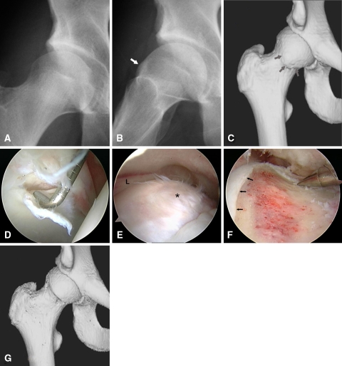
Fig. 2A–G.
Images illustrate the case of a 20-year-old hockey player with a 4-year history of right hip pain. (A) An anteroposterior radiograph is unremarkable. (B) A frog lateral radiograph demonstrates a morphologic variant with bony buildup at the anterior femoral head-neck junction (arrow) characteristic of cam impingement. (C) A 3-D CT scan further defines the extent of the bony lesion (arrows). (D) Viewing from the anterolateral portal, the probe introduced anteriorly displaces an area of articular delamination from the anterolateral acetabulum characteristic of the peel back phenomenon created by the bony lesion shearing the articular surface during hip flexion. (E) Viewing from the peripheral compartment, the bony lesion is identified (*) immediately below the free edge of the acetabular labrum (L). (F) The lesion has been excised, recreating the normal concave relationship of the femoral head-neck junction immediately adjacent to the articular surface (arrows). Posteriorly, resection is limited to the midportion of the lateral neck to avoid compromising blood supply to the femoral head from the lateral retinacular vessels. (G) A postoperative 3-D CT scan illustrates the extent of bony resection. (Reprinted with permission from J. W. Thomas Byrd, MD.)

