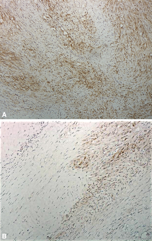
Fig. 1A–B.
Immunohistologic micrographs of a sample of a Dupuytren’s nodule show (A) an involutional zone with partially aligned staining myofibroblasts (larger spindle-shaped cells) with few fibers (Patient 1-8) and (B) an involutional β-catenin-staining zone (black arrow) adjacent to a nonstaining residual zone (white arrow) (Patient 1-12) (Stain, primary antibody for β-catenin; original magnification, ×400).

