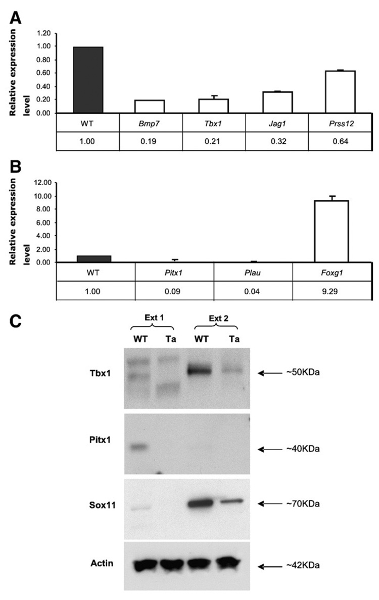
Fig. 2. Relative expression levels for selected genes in Ta primary keratinocytes.
(A) Bmp7, Tbx1, Jag1 and Prss12 from the “candidate EDA target” group showed significant downregulation in Ta keratinocytes, with WT set to 1.0. (B) Pitx1, Plau and Foxg1 from the “preliminary candidate” group also showed significant expression changes that were consistent with microarray results. (C) Western blotting assays showed that Tbx1 protein was significantly downregulated in Ta keratinocytes in the Ext 2 fraction (see Materials and methods). Pitx1 and Sox11 were also downregulated in Ta keratinocytes in Ext 1 and Ext 2 fractions, respectively. Actin was a loading control.

