
Fig. 1.
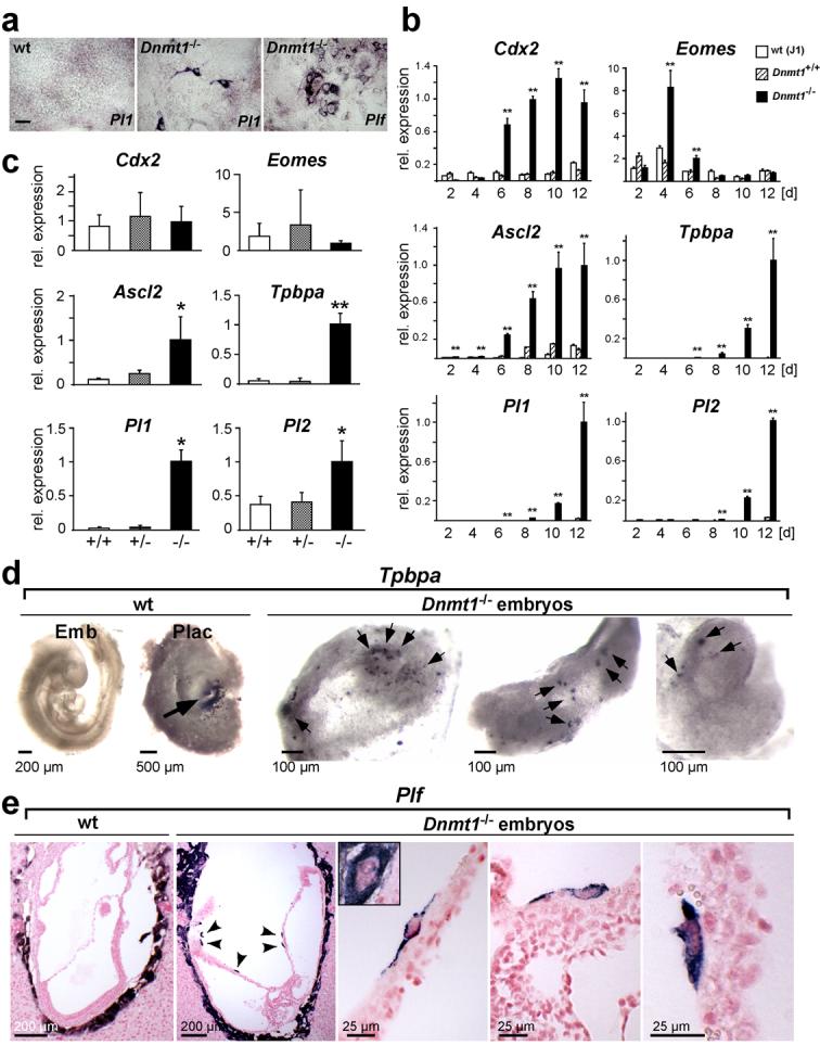
Dnmt1-deficiency enables trophoblast differentiation from cells committed to the embryonic cell lineage.
(a) Trophoblast giant cells differentiate from Dnmt1-/- ES cells, but not wildtype (wt) ES cells, in vitro when cultured in TS cell conditions. In situ hybridization with trophoblast giant cell-specific markers Pl1 (after 12d) and Plf (after 8d). (b) Time course of trophoblast marker activation in ES cells upon culture in TS cell medium for 2-12 days. Significant expression levels are only observed in Dnmt1-deficient ES cells (data are mean ± s.d., **P<0.005; n=3). Trophoblast stem cell markers (Cdx2, Eomes) are the first to be up-regulated, followed by intermediate diploid trophoblast markers Ascl2 and Tpbpa, and trophoblast giant cell markers Pl1 and Pl2. (c) Quantitative RT-PCR analysis of trophoblast markers on E9.5 embryos from Dnmt1+/- intercrosses. Trophoblast-restricted genes Ascl2, Tpbpa, Pl1 and Pl2 are detected at significant levels only in Dnmt1-/- embryos (data are mean ± s.d., *P<0.05, **P<0.005; n=7). (d) Whole mount in situ hybridization with the trophoblast marker Tpbpa on E9.5 Dnmt1-/- embryos and littermate control (wt) embryos and placentas. Positive staining (dark purple-black) is seen in the ectoplacental cone/spongiotrophoblast area (arrow) of the placenta (Plac). No staining is observed in the wt embryo (Emb). In Dnmt1-/- embryos, numerous Tpbpa-positive cells are present (arrows). (e) In situ hybridization with the giant cell marker Plf on E8.5 wildtype (wt) and Dnmt1-deficient conceptuses. Arrowheads point towards Plf-positive cells (blue-purple) that are observed within embryonic structures in Dnmt1-/- conceptuses only. Higher magnification views show the morphology of these cells; they are larger than surrounding cells but do not reach the size of parietal trophoblast giant cells (shown in inset). Scale bar in a represents 100 μm, scale bars in d and e are as indicated.