
Figure 7. Kinase client association with HSP90 and proteasomal degradation after silencing of CDC37 and 17-AAG.
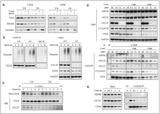
(a) Separation of proteins into total, soluble, or detergent-insoluble fractions after 72h siRNA transfection of human HCT116 colon cancer cells followed by treatment with DMSO, MG132 or 17-AAG or drug combination at 5XGI50 concentrations. (b) Left: Ubiquitin from CDK4 or goat IgG immunoprecipitation of total lysate after siRNA transfection for 72h and 0, 8 or 16h MG132 treatment at 5XGI50, right: input from total proteins. (c) CDK4 immunoprecipitates from pulse chase, labelling for 2h with 35S Met/Cys protein labelling mix after 96h siRNA transfection. Ctl is without antibody. The immunoprecipitates were also analysed by western blotting (WB). Note that the incorporation of the label is similar in CDC37 silenced versus non-silenced cells and that the turnover of newly synthesised CDK4 is comparable with both conditions (t1/2 ∼8h). (d) CDK4 immunoprecipitation following 24h 17-AAG treatment after siRNA or mock transfection for 72h. HSP90, HSC70, CDC37 and cyclin D1 association with CDK4. (e) Immunoprecipitation of CDK4 after 30min, 1h, 2h or 4h 17-AAG treatment at 5XGI50 and association with HSP90 and CDC37, Ctl IP is without antibody.