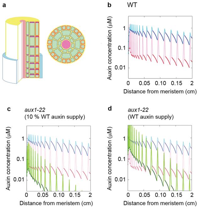
Figure 4. Expression of AUX1 in non-hair cells affects the supply of auxin to hair cells in the differentiation zone of the root.

(a) The left-hand image shows a sketch of the computer model of the Arabidopsis root apex. The epidermis, cortex, and endodermis are resolved in the elongation and differentiation zones. Auxin is supplied to the tissue by the proximal end of the lateral root cap (blue) and transported basipetally by the activity of AUX1 influx carriers (orange) and PIN family efflux carriers (plum). Cell membranes without influx carriers are shown in grey. Auxin is free to diffuse through the apoplast (yellow), which forms a continuous compartment between cells, and through the cytoplasm of cells (green). The right-hand image shows a transverse section through the model root showing cell geometry. Cells overlying a junction between two cortical cells are hair (H) cells; cells that do not overly a junction are non-hair cells (N). AUX1 expression is limited to N cells. (b) Model results showing the concentration of auxin in N cell files (blue) and H cell files (red) over increasing distance from the root tip. Dark colours show cytoplasmic auxin concentration, lighter colours show the apoplastic concentration between cells in a longitudinal file. (c) Model results for the aux1 mutant (green) superimposed on the wild-type results from panel b, assuming auxin supply from the cap is 10 % of wild-type. (d) Model results for the aux1 mutant assuming that the auxin supply from the lateral root cap is unchanged from wild type.
