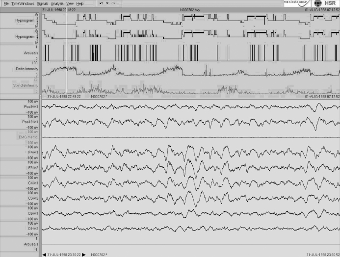
Figure 1.
Single case 1: 24-year-old man (healthy subject) who shows “no changes” in the sleep scoring parameters. In addition to the R&K and AASM hypnograms (first and second traces, respectively), arousals, delta and spindle intensity as revealed by the Somnolyzer 24×7 adapted for the AASM standard are shown in the upper part of the figure. The lower part shows a typical 30-s epoch in sleep stage N3 with 2 EOG derivations (Pos8-M1 and Pos18-M1), a mental EMG derivation, and 6 EEG derivations (F4-M1, F3-M2, C4-M1, C3-M2, O2-M1, O1-M2), as well as a channel indicating automatically detected arousals. To facilitate comparisons, all scales are the same for Figures 1 to 3. Note that the high-amplitude slow waves surpass the 75 μV criteria by far already at central leads.

