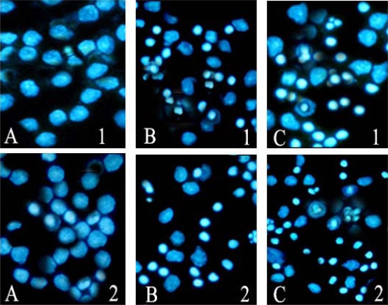
Figure 5.
Apoptosis observed by Hoechst 33258 staining (200×). After the cells were treated with 40 μmol/L ponicidin, Hoechst 33258 staining was used to observe the morphological changes of cell apoptosis. Morphological changes of cell apoptosis such as condensation of chromatin and nuclear fragmentations were found clearly. A: Control; B: Cells treated for 48 h; C:Cells treated for 72 h. 1. U937; 2. THP-1.

