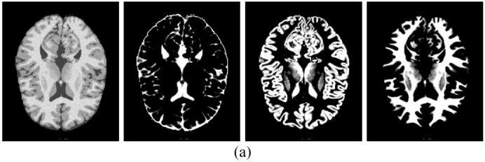
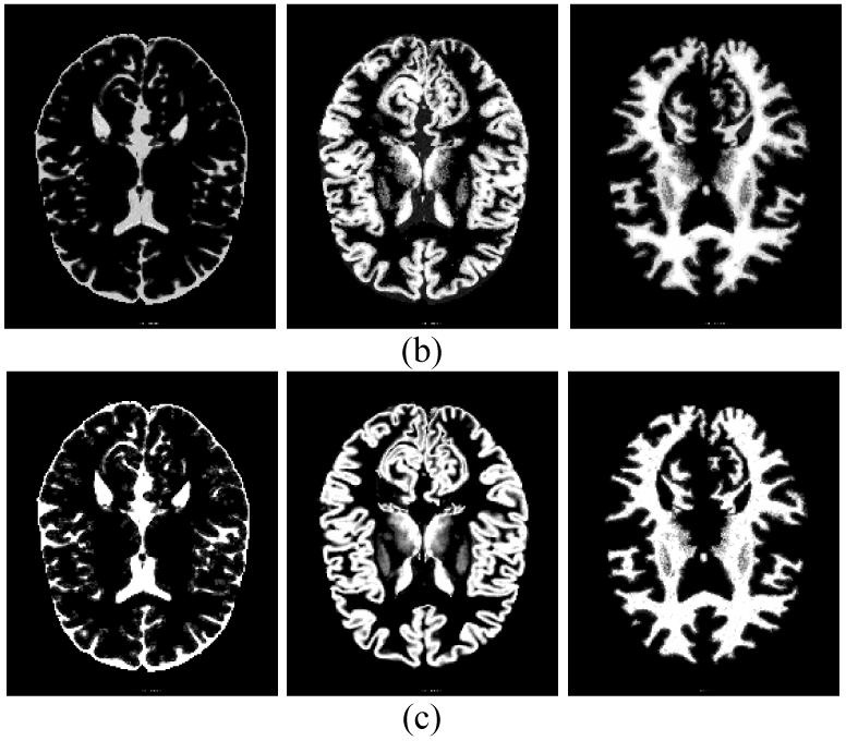
Fig. 7.


Simulated MRI brain phantom: (a) from left to right, they are noise phantom image at the 82nd slice, ground truth of {Zi1} for CSF, ground truth {Zi2} for grey matter, and ground truth {Zi3} for white matter respectively, (b) estimated {Zik} by the theoretical MAP-EM solution, and (c) estimated {Zik} by the approximated MAP-EM solution.
