Figure 3a.

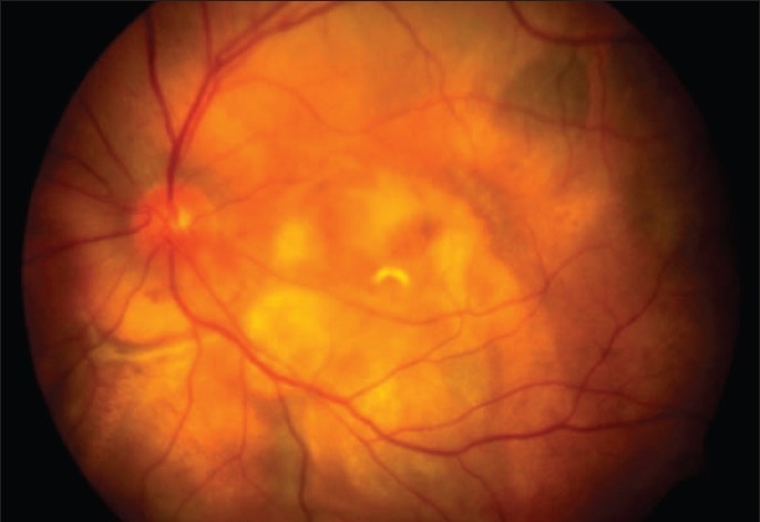
a: Color fundus photograph of the left eye showing the presence of scarring with surrounding hyperpigmentation in the area of the earlier mass, in the macular region and extending below the inferotemporal vascular arcade

a: Color fundus photograph of the left eye showing the presence of scarring with surrounding hyperpigmentation in the area of the earlier mass, in the macular region and extending below the inferotemporal vascular arcade